Welcome!

Hello, and welcome to our site about bacteria cell function and structure. We are KGDMX, and we hope to help you understand more about bacteria. Take a look at the side bar and learn fun, interesting facts at our fun facts section and also click on the quick links to learn more about the various topics available here.
Enjoy your time at our website! :D

- KGDMX

Friday, July 31, 2009

Pili and Fimbriae


Written By: Xiu Li
MB0902
092908H
A pilus is a hairlike appendage found on the surface of many bacteria. The term pilus and fimbriae are used interchangeably, although some researchers reserve the term pilus for the sexual appendage required for bacterial conjugation.

Pili connect a bacterium to another of its species, or to another bacterium of a different species, and build a bridge between the cytoplasm of the cells. This enables the transfer of plasmids between the bacteria, an exchanged plasmid can code for new functions. The pilus is made out of the protein flagellin.
Up to ten of these structures can exist on the bacteria. Some bacterial viruses or bacteriophages attach to receptors (a protein molecule embedded in either the plasma cembrane or cytoplasm of a cell) on sex pili at the start of their cycle.


Sex pili

A pilus is typically 6 to 7nm in diameter.


Despite its name, the sex pilus is not used for sexual reproduction.
During bacterial conjugation, a sex pilus emerging from one bacterium ensnares the recipient bacterium, draws it in, and eventually triggers the formation of a mating bridge. This establishes direct contact, merging the cytoplasms of two bacteria via a controlled pore. This pore allows for the transfer of bacterial DNA from the bacteria with the pilus (donor) to the recipient bacteria.


Through this mechanism of genetic transformation, advantageous genetic traits can be disseminated amongst a population of bacteria. Not all bacteria have the ability to create sex pili, however sex pili can form between bacteria of different species.


The fertility factor is required to produce a sex pili.
(A bacterial DNA sequence that allows a bacterium to produce a sex pilus necessary for conjugation)


Fimbriae


A fimbria is a a proteinaceous appendage in many gram-negative and gram-positive bacteria that is thinner and shorter than a flagellum. This appendage ranges from 3-10 nanometers in diameter and can be up to several micrometers long. In Gram positive bacteria, the pilin subunits are covalently linked.

Fimbriae are used by bacteria to adhere to one another and to adhere to animal cells, and some inanimate objects. Attachment of bacteria to host surfaces is required for colonization during infection or to initiate formation of a biofilm. Mutant bacteria that lack fimbriae cannot adhere to their usual target surfaces and, thus, cannot cause diseases.

Fimbriae are either located at the poles of a cell, or are evenly spread over its entire surface. A bacterium can have as many as 1,000 fimbriae.

Fimbriae are only visible with the use of an electron microscope.

Bacterial Cell Wall structure & Chemistry


Written By: Ginette
MB0902
092184Q


Composition & Characteristics


Component:
-Petidoglycan

Peptidoglycan, which is also known as murein, is a polymer consisting of sugars and amino acids that forms a mesh-like layer outside the plasma membrane of bacteria, forming the cell wall. The sugar component consists of alternating residues of β-(1,4) linked N-acetylglucosamine and N-acetylmuramic acid residues. Attached to the N-acetylmuramic acid is a peptide chain of three to five amino acids. The peptide chain can be cross-linked to the peptide chain of another strand forming the 3D mesh-like layer. Some Archaea have a similar layer of pseudopeptidoglycan or pseudomurein, where the sugar residues are β-(1,3) linked N-acetylglycosamine and N-acetyltalosaminuronic acid That is why the cell wall of Archaea is insensitive to lysozyme.

Functions:

  • Maintains characteristic shape of cell

  • Prevents cell from bursting

  • Virulence-ability to cause disease
  • Differentaiting between types of bacteria (Gram positive and negative)


Gram-positive bacteria

  • cytoplasmic lipid membrane
  • thick peptidoglycan layer(60%to 90%)

-teichoic acids and lipoids are present, forming lipoteichoic acids which serve to act as chelating agents, and also for certain types of adherence.

  • capsule polysaccharides (only in some species)
  • flagellum (only in some species)

-if it is present, it contains two rings for support as opposed to four in Gram-negative bacteria because Gram-positive bacteria have only one membrane layer.

Gram-negative bacteria

  • Cytoplasmic membrane
  • Thin peptidoglycan layer (10% to 20%)
  • Outer membrane contains lipopolysaccharide (LPS, which consists of lipid A, core polysaccharide, and O antigen) outside the peptidoglycan layer
  • Porins which are proteins that permit small molecules exist in the outer membrane
  • Periplasmic space: A space between the layers of peptidoglycan and the secondary cell membrane (contains digestive enzymes and transport proteins)
  • No teichoic acids or lipoteichoic acids are present
  • Lipoproteins are attached to the polysaccharide backbone.
  • Most do not sporulate (Coxiella burnetii, which produces spore-like structures, is a notable exception)

Differences between Gram postive and Gram negative Bateria Cell Walls
Peptidoglycan -Thick layer in Gram positive; thin layer in Gram negative
Teichoic acid - Present in Gram positive; Absent in Gram negative
Lipids -Very little in Gram positive; LPS in Gram negative
Outer membrane - Absent in Gram positive; Present in Gram negative
Periplasmic space - Absent in Gram positive; Present in Gram negative

Gram Staining
It is use to differentiate Gram-positive and Gram-negative bacteria based on the chemical and physical properties of their cell walls. Gram staining is not used to classify archaea, since these microorganisms yield widely varying responses that do not follow their phylogenetic groups.
Reagants:

  • Crystal violet (Primary Stain)
  • Iodine (Mordant)
  • Alcohol (Decolourizer)
  • Safrain ( Counter Stain)

Crystal violet (CV) dissociates in aqueous solutions into CV+ and chloride (Cl – ) ions. These ions penetrate through the cell wall and cell membrane of both Gram-positive and Gram-negative cells. The CV+ ion interacts with negatively charged components of bacterial cells and stains the cells purple. Iodine (I – or I3 – ) interacts with CV+ and forms large complexes of crystal violet and iodine (CV – I) within the inner and outer layers of the cell. When a decolorizer such as alcohol or acetone is added, it interacts with the lipids of the cell membrane. A Gram-negative cell will lose its outer membrane and the peptidoglycan layer is left exposed. The CV – I complexes are washed from the Gram-negative cell along with the outer membrane. In contrast, a Gram-positive cell becomes dehydrated from an ethanol treatment. The large CV – I complexes become trapped within the Gram-positive cell due to the multilayered nature of its peptidoglycan. The decolorization step is critical and must be timed correctly; the crystal violet stain will be removed from both Gram-positive and negative cells if the decolorizing agent is left on too long (a matter of seconds).
After decolorization, the Gram-positive cell remains purple and the Gram-negative cell loses its purple color. Counterstain, which is usually positively-charged safranin or basic fuchsin, is applied last to give decolorized Gram-negative bacteria a pink or red color.

Atypical Cells Walls
1. Archaea
- Extreme environmental conditions: methanogens, halophiles, thermophiles
- May lack cell wall
- Walls : No peptidoglycan, have pseudomurein

2. Mycoplasma
- Smallest bacteria (pass through filters for bacteria)
- Very little or no cell wall (very weak to ouside environment)
- Can withstand osmotic lysis

(plasma membrane:sterols, carotenoids, lipoglycans) =>confers strength

References:

http://en.wikipedia.org/wiki/Peptidoglycan

http://en.wikipedia.org/wiki/Gram-positive_bacteria

http://en.wikipedia.org/wiki/Gram-negative_bacteria

http://en.wikipedia.org/wiki/Gram_staining

http://en.wikipedia.org/wiki/Archaea

http://en.wikipedia.org/wiki/Mycoplasma

Nucleoid


Written By: Doraline
MB0902
092472X
The nucleoid is an irregularly-shaped region within the cell of prokaryotes which has nuclear material without a nuclear membrane and where the genetic material is localized. The genome of prokaryotic organisms generally is a circular, double-stranded piece of DNA, of which multiple copies may exist at any time. The length of a genome widely varies, but generally is at least a few million base pairs. Storage of the genome within a nucleoid can be contrasted against that within eukaryotes, where the genome is packed into chromatin and sequestered within a membrane-enclosed organelle called the nucleus.


Source from: http://en.wikipedia.org/wiki/File:Celltypes.png


A genophore is the DNA of a prokaryote, commonly referred to as a prokaryotic chromosome. The term chromosome is misleading for a genophore because the genophore lacks chromatin. The genophore is compacted through a mechanism known as supercoiling, where a chromosome is compacted via chromatin. The genophore is circular in most prokaryotes, and linear in very few. The circular nature of the genophore allows replication to occur without telomeres. Genophores are generally of a much smaller size than Eukaryotic chromosomes. Many eukaryotes carry genophores in organelles such as mitochondria and chloroplasts. These organelles are very similar to true prokaryotes.


The nucleoid can be clearly visualized on an electron micrograph at high magnification, although its appearance may differ, it is clearly visible against the cytosol. Sometimes even strands of what is thought to be DNA are visible. By staining with the Feulgen stain, which specifically stains DNA, the nucleoid can also be seen under a light microscope.

source from:http://biology.kenyon.edu/courses/biol114/Chap01/chrom_struct.html


Other links / references for nucleoid:
http://student.ccbcmd.edu/courses/bio141/lecguide/unit1/prostruct/nucleoid.html
http://www.reference.com/browse/wiki/Nucleoid

Flagellum


Written By: Xiu Li
MB0902
092908H


A Flagellum is a tail-like structure that projects from the cell of certain cells. In this post, I'll be explaining Bacterial Flagella, as shown by the picture above.
Flagella are made up of the protein flagellin and its shape is a 20 nanometer-thick hollow tube. They are helical filaments that have a sharp bend just outside the outer membrane, functions in locomotion and rotate like screws. The sharp bend, or "hook", allows the helix to point directly away from the cell.


A shaft runs between the hook and the basal body, passing through protein rings in the cell's membrane that act as bearings. Gram-positive organisms have 2 of these basal body rings, one in the peptidoglycan layer and one in the plasma membrane. Gram-negative organisms have 4 such rings: the L ring associates with the lipopolysaccharides, the P ring associates with peptidoglycan layer, the M ring is embedded in the plasma membrane, and the S ring is directly attached to the plasma membrane. The filament ends with a capping protein.


Video! : http://www.youtube.com/watch?v=0N09BIEzDlI

The bacterial flagellum is driven by a rotary engine made up of protein located at the flagellum's anchor point on the inner cell membrane. The engine is powered by proton motive force, i.e., by the flow of protons (hydrogen ions) across the bacterial cell membrane due to a concentration gradient set up by the cell's metabolism. The rotor transports protons across the membrane, and is turned in the process.


Flagella do not rotate at a constant speed but instead can increase or decrease their rotational speed in relation to the strength of the proton motive force. Flagellar rotation can move bacteria through liquid media at speeds of up to 60 cell lengths/second (sec). Although this is only about 0.00017 km/h (0.00011 mph), when comparing this speed with that of higher organisms in terms of number of lengths moved per second, it is extremely fast


The flagellar filament is the long helical screw that propels the bacterium when rotated by the motor, through the hook. In most bacteria that have been studied, the filament is made up of eleven protofilaments approximately parallel to the filament axis. Each protofilament is a series of tandem protein chains.


Through use of their flagella, bacteria who have them are able to move rapidly towards attractants and away from repellents. They do this by means of a Biased Random Walk (in which bacteria search for food or flee from harm), with 'runs' and 'tumbles' brought about by rotating the flagellum


Flagella arrangement schemes


Different species of bacteria have different numbers and arrangements of flagella. There are basically four different types of flagellar arrangements.


1. A single flagellum can extend from one end of the cell - if so, the bacterium is said to be monotrichous.
2. A single flagellum (or multiple flagella) can extend from both ends of the cell - amphitrichous.
3. Several flagella (tuft) can extend from one end or both ends of the cell - lophotrichous; or,
4. Multiple flagella may be randomly distributed over the entire bacterial cell - peritrichous.

(amphi- a prefix meaning both or on both sides
lopho- or loph- a combining form meaning a "ridge" or "tuft,")


In some bacteria, the individual flagella are organized outside the cell body, helically twining about each other to form a thick structure called a fascicle. Other bacteria have a specialized type of flagellum called an "axial filament" that is located in the periplasmic space, the rotation of which causes the entire bacterium to move forward in a corkscrew-like motion.


Counterclockwise rotation of monotrichous polar flagella pushes the cell forward with the flagella trailing behind, much like a corkscrew moving inside cork. The flagella are left-handed helices, and bundle and rotate together only when rotating counterclockwise. When some of the rotors reverse direction, the flagella unwind and the cell starts "tumbling".
Such "tumbling" may happen occasionally, leading to the cell seemingly thrashing about in place, resulting in the reorientation of the cell. The clockwise rotation of a flagellum is suppressed by chemical compounds favorable to the cell (e.g. food), but the motor is highly adaptive to this. Therefore, when moving in a favorable direction, the concentration of the chemical attractant increases and "tumbles" are continually suppressed; however, when the cell's direction of motion is unfavorable (e.g., away from a chemical attractant), tumbles are no longer suppressed and occur much more often, with the chance that the cell will be thus reoriented in the correct direction.


(arrows are pointing to the Flagella)

References: http://en.wikipedia.org/wiki/Flagellum
http://www.life.umd.edu/classroom/bsci424/BSCI223WebSiteFiles/Flagella.htm

Thursday, July 30, 2009

Cytoplasm


Written By: Michelle
MB0902
091715M

What is cytoplasm?

  • A viscous gel inside the membrane. It liquifies when shaken or stirred.
  • It is the part of a cell that is enclosed within the plasma/cell membrane.
  • The cytoplasm is the site where most cellular activities occur, such as many metabolic pathways like glycolysis, and processes such as cell division.

What does it contains?

  • Contains a lot of water, and the other organelles of the cells.
  • It is made up of proteins, vitamins, ions, nucleic acids, amino acids, sugars, carbohydrates and fatty acids.
All of the functions for cell expansion, growth and replication are carried out in the cytoplasm of a cell.


Some reference links:
http://www.essortment.com/all/cytoplasm_rkkg.htm
http://en.wikipedia.org/wiki/Cytoplasm
http://sln.fi.edu/qa97/biology/cells/cell3.html

Ribosomes


Written By: Doraline
MB0902
092472X

Ribosomes are small organelles made of rRNA and protein in the cytoplasm of prokaryotic and eukaryotic cells which aid in the production of proteins on the rough endoplasmic reticulum and ribosome complexes. The site of protein synthesis. Ribosomes that are from bacteria, archaea and eukaryotes have significantly different in its structure and it’s RNA.
The ribosome is composed of two subunits that attach to the mRNA at the beginning of protein synthesis in the cytoplasm and detach when the polypeptide has been translated. Ribosomal subunits are synthesized by the nucleolus.


(Blue-large subunit. Red- Small subunit)


The ribosome functions in the expression of the genetic code from nucleic acid into protein, in a process called translation. Ribosomes do this by catalyzing the assembly of individual amino acids into polypeptide chains. This involves the binding of a mRNA and then using this as a template to join together the correct sequence of amino acids. This reaction uses adapters called transfer RNA molecules, which reads the sequence of the mRNA and are attached to the amino acids.


There are two places that ribosomes usually exist in the cell:
1. suspended in the cytosol
2. bounded to the endoplasmin reticulum.
These ribosomes are called free ribosomes and bound ribosomes respectively. In both cases, the ribosomes usually form aggregates called polysomes.Free ribosomes usually make proteins that will function in the cytosol, while bound ribosomes usually make proteins that are exported or included in the cell's membranes. Interestingly enough, free ribosomes and bound ribosomes are interchangeable and the cell can change their numbers according to metabolic needs!!


Prokaryotes have 70S ribosomes, each consisting of a small (30S) and a large (50S) subunit. Their large subunit is composed of a 5S RNA subunit (consisting of 120 nucleotides), a 23S RNA subunit (2900 nucleotides) and 34 proteins. The 30S subunit has a 1540 nucleotide RNA subunit (16S) bound to 21 proteins.
Eukaryotes have 80S ribosomes, each consisting of a small (40S) and large (60S) subunit. Their large subunit is composed of a 5S RNA (120 nucleotides), a 28S RNA (4700 nucleotides), a 5.8S subunit (160 nucleotides) and ~49 proteins. The 40S subunit has a 1900 nucleotide (18S) RNA and ~33 proteins.


Source :http://en.wikipedia.org/wiki/Ribosome
(The image shows an isosurface representation of the 70S ribosome (50S blue; 30S yellow), derived from a crystal structure of E. coli ribosome - Schuwirth et al (2005).)


Source: from http://www.denniskunkel.com/


Other Link / references about ribosomes:
http://www.answers.com/ribosomes
http://www.stemnet.nf.ca/~dpower/cell/ribo.htm
http://www.daviddarling.info/encyclopedia/R/ribosome.html
http://www.youtube.com/watch?v=Jml8CFBWcDs (video)

Plasma Membrane


Written By: Michelle
MB0902
091715M

Scope:

  • 1. Functions and composition of cell membrane.
  • 2. Selective permeability.
  • 3. Types of movements - Passive and Active.

1. Functions and Composition of Cell Membrane.
The cell membrane (also called the plasma membrane or plasmalemma) is the biological membrane separating the interior of a cell from the outside environment.

Roles of cell membrane:

  • - It encloses the cytoplasm.
  • - The cell membrane also plays a role in anchoring the cytoskeleton to provide shape to the cell.


Fig 1.

The cell membrane is composed of:

  • - Phospholipid (PL) Bilayer, both hydrophilic and hydrophobic.
  • - Proteins - interspersed between the phospholipid bilayer and acts as pores/channels in movement of materials in/out of the cell.

Phospholipids are amphiphilic with the hydrocarbon tail of the molecule being hydrophobic; its polar head hydrophilic.
As the plasma membrane faces watery solutions on both sides, its phospholipids accommodate this by forming a phospholipid bilayer with the hydrophobic tails facing each other.

Fig 2.

2. Selective Permeability

  • - Allows only certain molecules and ions in and out of the cell.
  • - Permeability depends on size and its permeability in lipids. See Fig 3.


Fig 3.

3. Types of Movements: Passive

Passive: Movements of materials from an area of high concentration to low concentration.

  • - No expenditure of Adenosine Triphosphate, ATP (energy).
  • - Further broken down into 3 types of movements:
  • A. Simple Diffusion.
  • B. Facilitated Diffusion.
  • C. Osmosis.

A. Simple Diffusion

Definition: Movement of substance from an area of higher concentration to lower concentration.
Movement will cease when the molecules or ions are equally distributed (equilibrium).

B. Facilitated Diffusion

Definition: Substances transported by transporter proteins across membranes from areas of high to low concentration.

Fig 4.

*Note: Always remember for simple and facilitated diffusion, molecules always move from HIGH to LOW. It's just remembering that for simple, it's the movement of substance whereas for facilitated, it's the transportation of substances. (:

C. Osmosis

Definition: It's the movement of water across a selective permeable membrane until equilibrium is reached.
Water molecules move from diluted to more concentrated area.

There are 3 types of osmotic solutions that bacteria may encounter:

  • Isotonic: equal number of water molecules
  • Hypotonic: water molecules entering the bacteria
  • Hypertonic: water molecules moving out of the bacteria

Fig 5.

3. Types of Movements: Active

Active: Movement of materials from an area of low concentration to high concentration.

  • - Expenditure of Adenosine Triphosphate ATP (energy).
  • - Further broken down into 2 types of movements:
  • A. Active Transport
  • B. Group Translocation

A. Active Transport

Definition: Substances transported by transporter proteins from low concentration to high concentration.
ATP is used during the process.

Fig 6.

B. Group Translocation

Definition: It is the use of energy such as phosphoenolpyruvic acid (PEP) to modify chemicals and transport them across the cell membrane.
Modified substances cannot leave the cells, this is useful when the cell is in an environment where nutrients are limited.

Stage 1: When bacteria use the process of group translocation to transport glucose across their membrane, a high-energy phosphate group from phosphoenolpyruvate (PEP) is transferred to the glucose molecule to form glucose-6-phosphate.

Stage 2: A high-energy phosphate group from PEP is transferred to the glucose molecule to form glucose-6-phosphate.

Stage 3: The glucose-6-phosphate is transported across the membrane.

Stage 4: Once the glucose has been converted to glucose-6-phosphate and transported across the membrane, it can no longer be transported back out.


That's about it.
Hope you guys can understand what I'm trying to convey about. (:

Some reference links:
http://en.wikipedia.org/wiki/Cell_membrane
http://en.wikipedia.org/wiki/Selective_permeability
http://academic.brooklyn.cuny.edu/biology/bio4fv/page/simple.htm
http://en.wikipedia.org/wiki/Facilitated_diffusion
http://en.wikipedia.org/wiki/Osmosis
http://en.wikipedia.org/wiki/Active_transport
http://en.wikipedia.org/wiki/PEP_group_translocation

Glycocalyx


Written By: Xiu Li
MB0902
092908H

Sugar Coat


That's literally what a Glycocalyx, of bacteria, is.
It's a network of polysaccharides (sugars) that project from cellular surfaces, such as bacteria. It serves to protect the bacterium by creating capsules, or allows the bacterium to attach itself to inert surfaces (like teeth or rocks), eukaryotes and even other bacteria! Their glycocalyxes can fuse together to envelop the colony.
There are two kinds of glycocalyxes. A distinct, gelatinous glycocalyx is called a Bacterial Capsule while an irregular, diffuse layer is called a Slime Layer.


Glycocalyx are made in cells and excreted to the cell surface, thus, they can be found just outside the cell wall of a bacterium. Being viscous (sticky), it helps in the formation of biofilms such as a coating on inert surfaces like catheters, teeth or rocks. This helps it in colonization as it resists flushing. Glycocalyx also helps protect bacteria from phagocytes (white blood cells), one way in which it does that is the resistance of phagocytic engulfment .
These traits increase the bacterium's virulence.

References: http://en.wikipedia.org/wiki/Glycocalyx http://student.ccbcmd.edu/courses/bio141/lecguide/unit1/prostruct/glyco.html


Sunday, July 26, 2009

Inclusions


Written By: Kiat Yi
MB0902
093712T

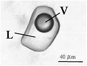
Cell Inclusions are aggregates of various compounds that are normally involved in storing energy reserves or building blocks for the cell. That means that they are made up of a combination of cell compounds, mainly proteins, and besides being the building blocks of the cell, they also store the cell’s excess energy for future use.


Cell inclusions are small, non-living intracellular (that means “inside the cell”) particles, usually represented as a form of stored food, and are not immediately vital to life processes. They also typically represent sites of viral multiplication in a bacterium or a eukaryotic cell and usually consist of viral capsid proteins (the protein shell of a virus).


Some examples of inclusions include:
  • metachromatic granules
  • polysaccharide granules
  • lipid inclusions
  • sulfur granules
  • carboxysomes
  • gas vacuoles

Reference Link: http://en.wikipedia.org/wiki/Inclusion_bodies

Endospores


Written by: Tan Kiat Yi
MB0902
093712T


Bacteria are vulnerable to damage from adverse environmental factors such as heat. So, there are a few types of bacteria that have a special trick up their “sleeve” that allows them to be dormant when the need arises. This “trick” is the ability to form resistant resting structures called endospores.

What they do
An endospore, as its name suggests, “endo” meaning “inside” and “spore” meaning “a resistant body”, is an internal structure formed by the bacterium that makes the bacteria dormant and tough. It ensures the survival of a bacterium through periods of environmental stress. Some examples of environments that endospores are capable of resisting are shown in the diagram below.
When the bacterium is put in favorable conditions again, it will revert to its vegetative state through germination, where it would carry out normal cell functions again. However, activation must take place first, it may be triggered by heating the endospore.

How they are formed

Endospores are formed through the process of sporulation. Under adverse environmental conditions, DNA replicates and a cytoplasmic membrane septum forms at one end of the cell. Another membrane then forms around one DNA molecule, and a forespore is formed. Then, the membrane layers synthesize peptidoglycan in the space between them to form the first protective coat, the cortex. A spore coat composed of a keratin-like protein then forms around the cortex. In some cases, an outer membrane made up of lipid and protein called an exosporium is seen. Finally, the remainder of the bacterium is degraded and the endospore is released. Sporulation usually takes around 15 hours.

Process of formation of endospore


Structure of endospore


In short, an endospore is an internal structure formed when the bacterium produces a thick internal wall that encloses its DNA and part of its cytoplasm.

Endospore forming bacteria
Not all bacteria can create endospores, only gram-positive bacteria can do so; some examples of endospore creating bacteria are the genus Bacillus, the genus Clostridium, and several other genera of bacteria including Desulfotomaculum, Sporosarcina, Sporolactobacillus, Oscillospira, and Thermoactinomyces. You can read more about gram-positive bacteria here.

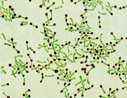
Endospores can be seen under the light microscope using only special staining techniques such as the Moeller stain and Schaeffer-Fulton stain, as its walls are impermeable to most dyes and stains.

Endospores stained green


Danger to Humans
Despite the few groups of endospore forming bacteria, people do usually come in contact with them. These bacteria can commonly be found in soil and water. Thankfully, most of these bacteria are harmless, and pose no threat to humans.
However, there are some important endospore producers that can cause serious infectious disease. Here are some brief examples of the above:

· Bacillus anthracis - the causative agent of anthrax

· Clostridium tetani - the agent of tetanus

· C. perfringens – may cause gas gangrene.

· C. botulinum – may cause botulism

· C. diff. – resistant to antibiotics

Destroying

Vaccinations and antibiotics are used to prevent and treat infections caused by endospore bacteria. However, these may prove pointless if the infection resides deep in the tissue. Also, even though endospores are resistant to extreme heat and radiation, they can be destroyed by burning or autoclaving, a process used to sterilize things. Exposure to extreme heat for an extended period of time, prolonged exposure to high energy radiation, such as x-rays and gamma rays would also affect

While resistant to extreme heat and radiation, endospores can be destroyed by burning or autoclaving. Exposure to extreme heat for a long enough period will generally have some effect, though many endospores can survive hours of boiling or cooking. Prolonged exposure to high energy radiation, such as x-rays and gamma rays, will also kill most endospores.

So there you have it, all you need to know about endospores. I hope that you've learned more about these amazing bacterial structures!

Here are some reference links:
http://en.wikipedia.org/wiki/Endospore
http://www.micro.cornell.edu/cals/micro/research/labs/angert-lab/bacterialendo.cfm
http://student.ccbcmd.edu/courses/bio141/lecguide/unit1/prostruct/spore.html
http://bacteriology.suite101.com/article.cfm/what_is_a_bacterial_endospore